Design
Because the project is all about the design
Principal of the design
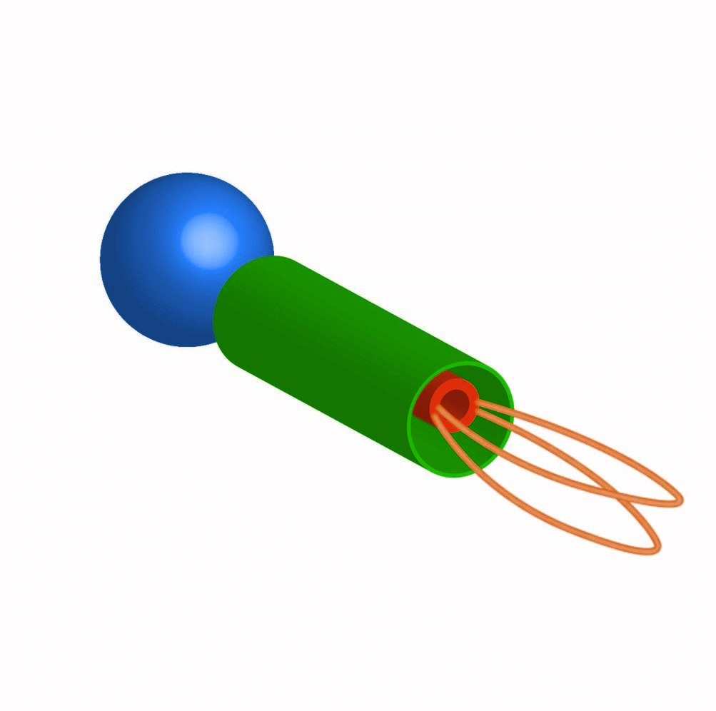
The design is fundamentally composed of an outer tube and an inner tube connected by several single strand DNA to keep the inner tube inside the larger outer tube. The inner tube has several protruding loops, serving as the part intended to capture particles. Streptavidin coated magnetic bead is then attached to the inner tube by having several protruding biotinylated DNA staples serving as attachment points to the magnetic bead. There are 2 designs that we created, a preliminary design, and a final design, both were designed using the software Cadnano.
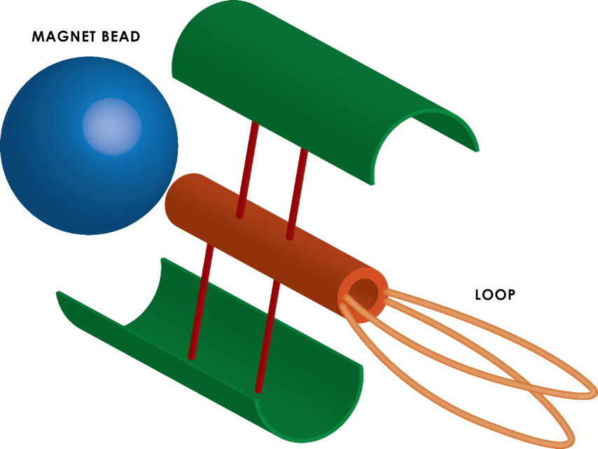
stranded DNAs connect the inner (orange) to the outer tube
(green), position and numbers are arbitrary.
Initial design
During Biomod Japan, we designed a structure with inner tube about 14nm in diameter and outer tube about 38nm in diameter,
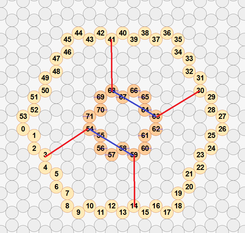
the red lines are the connections and the blue lines
are the places where the loops are going to connect.
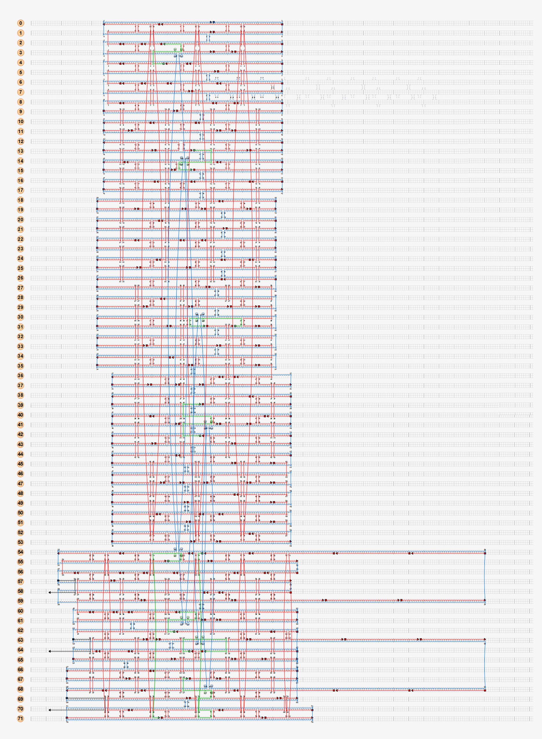
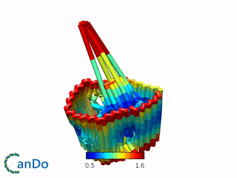
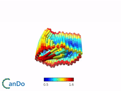
Fig 2.5. Simulation by Cando.
Problems of the first design
This design created several problems regarding stability as the skeleton of the tubes have a relatively bigger diameter. Moreover, observation using FRET will be hard due to the distortion of the tubes and the movement of the inner tube relative to the larger tube. (link to FRET). Finally, the relatively big size of this structure meant that there was not really any space to modify the length of the structure to be longer to accommodate as the m13mp18 single strand DNA has a total length of 7149 base pairs.
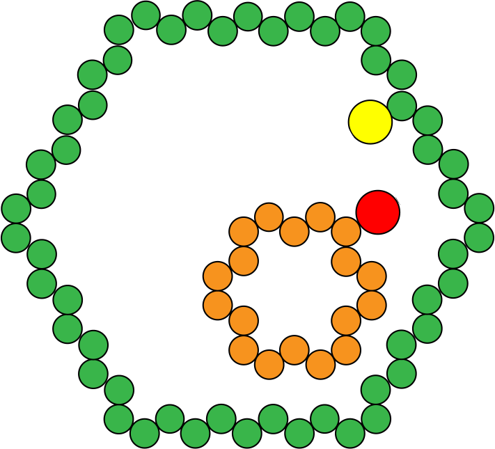
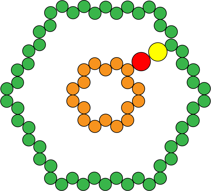
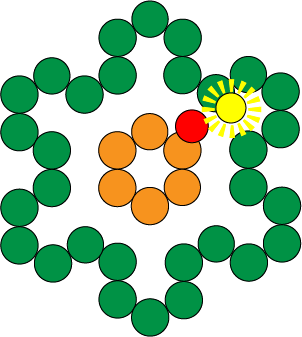
Fig 2.6. Movement of inner tube in relative to outer tube (left) can be minimized by sizing down both of the tubes (right).
final design
As the second and final structure, we proposed a design that was smaller in diameter, this made it more feasible to conduct observations such as FRET as the movement of the inner tube relative to the outer tube can be regulated. Therefore, it also meant that the whole structure is far more stable and movement due to distortion of the tubes can be minimized, this was done by adjusting the diameter of the inner tube to 6nm and the outer tube to about 18nm. The connections between the inner and outer tube were moved to outside of the tube to not hinder the sliding movement of the inner tube. The length of the structure including the loop is going to be about 109nm, and the outer tube will be about 50nm. The sequences of the strands can be downloaded from here
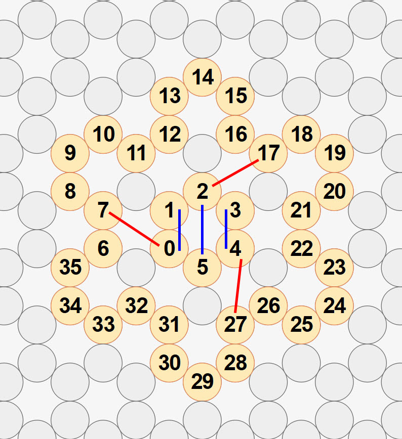
Fig 2.7. top view of the structure as seen from cadnano. The blue lines are where the loops connect and the red lines are the connections between the outer and smaller tube.
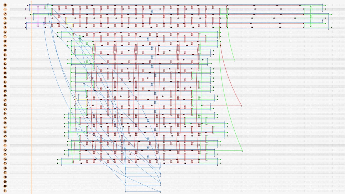
Fig 2.8. cadnano view of the structure.
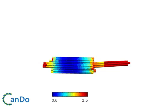
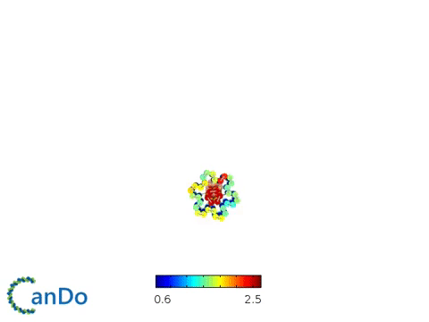
fig 2.9. Cando simulation of the final structure.
3 Staples from the inner tube have ends modified with biotin to provide means of attachment to the streptavidin coated magnetic bead. The sequence is as following:
| Name | Sequence |
|---|---|
| Biotin (199) | Biotin 5’ TA AAA GAA GTT TTT TCC AGA CGT TTT 3’ |
| Biotin (198) | Biotin 5’ TT TTT TAG TAA ATG AAT TTT GTC GTC TGC CAG AG 3’ |
| Biotin (200) | Biotin 5’ TT TTT TTT AGA GGC GGT TTG AAT AGC GAG AGG CTT TTG CTT TT 3’ |
Table 1. Sequence of the staples
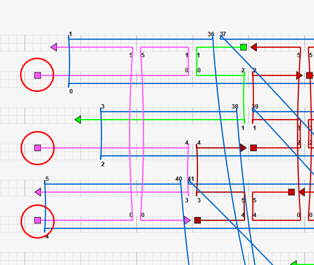
Fig 2.10. Staples modified with biotin as seen from cadnano (circled red).